CT SCAN
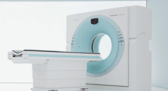
iRIS Imaging & Diagnostics is proud to announce that we are the first center in the surrounding areas of Vijayawada that uses a state-of-the-art Siemens sensation 64-slice Computed Tomography (CT) Machine with Ultra Low Dose Radiation Technology in our spacious premises. We offer advanced diagnostic services, facilitated by our exceptional team of Radiologists with immediate reporting at affordable prices for our patients.

What Is A CT (Computed Tomography) Scan?
A Computed Tomography (CT) scan is a sophisticated diagnostic tool that uses X-Ray technology and a high-speed computer to capture images in-detail and study the various parts and organs of your body such as
Chest
Stomach
Pelvis
Limbs
Heart
Liver
Lungs
Kidney
Adrenal Glands
Bladder
Pancreas
Intestines
Blood vessels
Bones
Spinal cord
We ask our patients to lie flat on a table that is attached to a large doughnut-shaped machine or the CT scanner. The scanner sends x-rays to the body parts or organs being studied.
Why Do We Use The Siemens Sensation 64-Slice CT Scan?
The Siemens 64-Slice CT Scan is an advanced, safe, and painless kind of diagnostic imaging which is usually recommended for the evaluation of soft tissue, such as the internal organs.
Let’s take the example of the heart. The heart is continually beating and changing its size and shape, and it’s tricky to get an accurate image of it, without causing blurring like other scanners. The Siemens 64-Slice CT Scan can capture a clear image of the heart and coronary vessels between each contraction in less than 5 seconds.
The Siemens 64-Slice CT Scan can identify cysts, tumour’s, and diseases of several organs such as the lung, liver, and coronary arteries. It can help visualize the blood flow in the arteries supplying to the various parts of the body, including the brain, lungs, kidneys and heart.
Some benefits of our Siemens 64-Slice CT Scan are:
Better view of the size, shape and position of the body part or organ
Superior image quality
Rapid scan time i.e. usually less than 15 seconds.
Lower dose of radiation
Better patient experience
Faster diagnostic time and results
Aids better treatment outcomes
The shortening of the time for scanning makes it ideal for patients who are children or those who cannot lie down in one position for a certain period.
How Does The Siemens 64-Slice CT Scan Work?
Unlike other standard scans, our low dose 64 slice CT x-ray source is rotated around the body parts or organs being studied. Each rotation of the CT scanner creates a cross-sectional image of the body organ or area called “slice” that happens simultaneously with the movement of the x-ray tube and table. This rotational movement ensures the maximum capture of images and information in a shorter time frame.
Each rotation creates 64 high-resolution anatomical images in less than a second. It takes less than 15 seconds for the complete scan. The slices are usually as thin as a half a millimeter or credit card. The multiple images captured at different angles during the rotations combine to form a three-dimensional view of the specified body part or organ. It helps the doctor get the most information when making any diagnosis.
Sometimes, we may ask patients to take in a contrast material orally, or via vein (IV) in their arm, or in other parts of their body such as a joint or rectum. We may also ask our patients to fast for some time before the scan. The contrast material looks white in the images to emphasize the specified body part or organ.
We may also take CT pictures ‘before and after’ using the contrast material.
Our CT scans are done by our experienced team of certified radiology technologists specialized in computed tomography. Our assigned technologist is always close by to assist our patients. The images are read by our on-site expert Doctors team of radiologists, who then sends the reports to the referring physician.