X-RAY
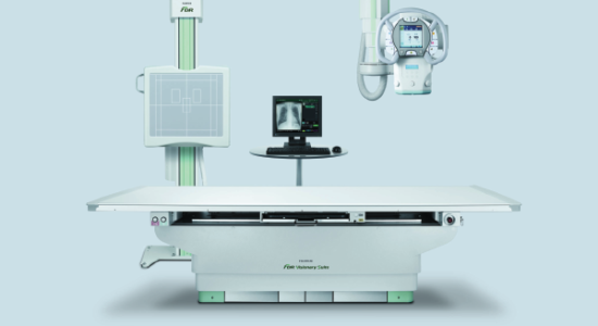
iRIS Imaging & Diagnostics is the first imaging center in the surrounding areas of Vijayawada to be equipped with FUJI Digital X-Ray that maximizes workflow efficiency and provides the best image quality with consistent reliability.

What Is An X-ray And Why Is It Used?
An X-ray is a quick, easy exam that is used to create images of your internal organs and bones using a small amount of radiation to help diagnose injuries and conditions.
Some of these health conditions include:
Arthritis
Fractures
Dislocations
Broken bones
Bone infections
Bone tumor’s
Kidney stones
Fluid in the lung
Hysterosalpingography
Barium meal
Foreign body location X-ray
Why iRIS Radiology Uses FUJI Digital X-Ray Technology?
Our FUJI Digital X-Ray scan helps physicians respond to health emergencies in sensitive environments safely and effectively with agile mobility.
Some beneficial features of the FUJI Digital X-Ray scan technology are:
Compact and lightweight.
Fast exam and cycle times.
Lesser patient’s waiting time.
Low dose radiation and power.
Portable and instant imaging.
Superior image quality.
Generous imaging area.
High volume caseloads.
Improved clinical outcomes.
Enhanced patient comfort, safety, and experience.
Suitable for routine diagnosis and complex trauma cases.
Improved infection controls with germ resistant surfaces.
What To Expect In Our X-Ray Scan At iRIS ?
Our courteous and experienced team at iRIS Imaging & Diagnostics will brief you before your scan about the preparation instructions, any special requirements, and your past imaging exams. You may be asked to bring your past imaging results with you when you come for the exam.
Our expert technologist will position you in our FUJI Digital X-Ray machine for the area of your body being picturized. Our technologist will go behind a partial wall with a viewing window to run the X-ray scan.
Some of our X-ray scans include fluoroscopy, a procedure which involves the administration of contrast orally, or an IV in your arm or hand, or into the area which is causing your pain. If your X-ray scan procedure requires an oral contrast, our team will inform you in advance during your appointment preparation call. You may experience a minor pressure sensation or temporary discomfort.
We use the fluoroscopy procedure as it helps guide the exact needle placement for injections to help treat pain. It aids in better diagnosis as it provides a view of the movement of internal anatomy or contrast material within the body. We use the fluoroscopy exam mainly for image-guided therapeutic injections into the joints or spine.
Our fluoroscopy procedure uses a continuous X-ray beam with a special machine called a C-arm that rotates around your body which projects a series of X-ray images captured at different angles onto a screen. The contrast material highlights the specific area and makes it possible for your doctor to view the internal organs and tissues in motion.
Depending on the complexity of your case, you may need additional imaging. If so, then you will be brought to a room where imaging will be performed via MRI, CT or X-ray scan.
Once your scan is complete, our team will escort you to the changing room so you can change into your clothing.
Our team will send your images electronically to our radiologist who will review the information and send a report to your referring healthcare provider, typically within X-Y business days.
You should follow up with your referring doctor to discuss your X-ray scan results.
Please make sure to inform us if you are pregnant, nursing, or might be expecting soon before you start the X-ray scan procedure. Although complications are rare in X-ray scans, our team will review the possible side effects and risks with your case prior to your exam. We will also ask certain questions to you and your referring doctor to decide if this X-ray exam is right for you.